• Skip to main content
  • Skip to primary sidebar
  • Skip to footer
  • Home
  • Insights & Articles
    • The Clinical Landscape: Strategic Insights into Retinal Care
      • The Modern Clinical Retina Landscape: A Strategic Overview
      • Surgical Approaches to Retinal Detachment: A Comparative Analysis
      • Gene Therapy in Retina: A Commercial and Clinical Appraisal
      • Evolving Treatment Paradigms for Diabetic Macular Edema
      • Analysis of the Anti-VEGF Market: From Revolution to Refinement
      • A Patient’s Guide to Understanding Age-Related Macular Degeneration (AMD)
    • The Business of Retina: Mastering Operations and Financial Health
      • Anatomy of a Retina Practice: A Strategic Guide to Financial Health and Operations
      • The Impact of Private Equity: Reshaping the Business of Retina
      • The Complete 2025 Guide to Ophthalmic J-Codes & Reimbursement
      • The Buy-and-Bill Model: A High-Risk, High-Reward Strategy
      • Staffing for Success: Key Roles in a High-Volume Retina Practice
      • Key Technology Investments for an Efficient Retina Clinic
      • Decoding CMS Reimbursement: A Guide to J-Codes and Revenue Cycle Management
    • The Innovation Pipeline: Shaping the Future of Retinal Care
      • Beyond the Needle: The Future of Drug Delivery in Retina
      • Heads-Up vs. Traditional: A Comparison of Surgical Visualization Platforms
      • How AI is Changing the Game in Retinal Disease Screening
      • The Rise of At-Home Monitoring for AMD
    • The Professional Ecosystem: Building a Career in Retina
      • A Day in the Life of a Retina Specialist: A Day of Preserving Sight
      • Industry vs. Academia: Choosing Your Career Path in Retina
      • The Role of Key Opinion Leaders (KOLs) in Retina
      • Maximizing Your ASRS Experience: A Fellow’s Guide
      • Navigating the Retina World: A Guide to Key Societies, Conferences, and Career Paths
  • Services
    • Search Engine Optimization (SEO) & Reputation Management
    • Content Marketing & Optometrist Network Development
    • Specialized Recruitment & Staffing Support
    • Professional Branding & Key Opinion Leader (KOL) Development
    • On-Demand Patient Education & Marketing Materials
    • Market Intelligence & M&A Consulting
  • Contact Us
    • Events
    • Subscribe
    • Submissions
Retina Consultant

Retina Consultant

Visionary News & Insightful Strategy

Retina Roundup Advertisement

Updated guidelines enhance choriocapillaris imaging in AMD with SS-OCTA

April 10, 2025 by Retina News Feed Leave a Comment

Click here to view the original post by Ophthalmology Times.

In addition to our own original articles, the Retina Consultant team expertly curates relevant news and research from leading publications.to provide our readers with a comprehensive view of the retina & ophthalmology landscape, Full credit belongs to the original authors and publication. 

By clicking the link above, you will be leaving our site to view an article from its original source.


Sharpen your perspective. Get our weekly analysis of the news shaping the retina industry.

Please enable JavaScript in your browser to complete this form.
Loading


Swept-source optical coherence tomography angiography (SS-OCTA) has become a powerful tool for imaging the choriocapillaris (CC) and showing that decreased CC perfusion plays an important role in the progression of age-related macular degeneration (AMD).1-4 Numerous studies have demonstrated that CC flow deficits increase with age and the CC is significantly impaired within the central macula, highlighting the potential link between CC dysfunction and AMD.5,6

SS-OCTA offers distinct advantages over spectral-domain OCTA (SD-OCTA) due to its longer wavelength, improved choroidal penetration, better image quality, and denser scan patterns but imaging the CC in AMD presents unique challenges, particularly when imaging areas affected by drusen and other pathological AMD changes such as soft drusen, calcified drusen (CaD), and hyperreflective foci (HRF).7-9

Advances in compensation strategies

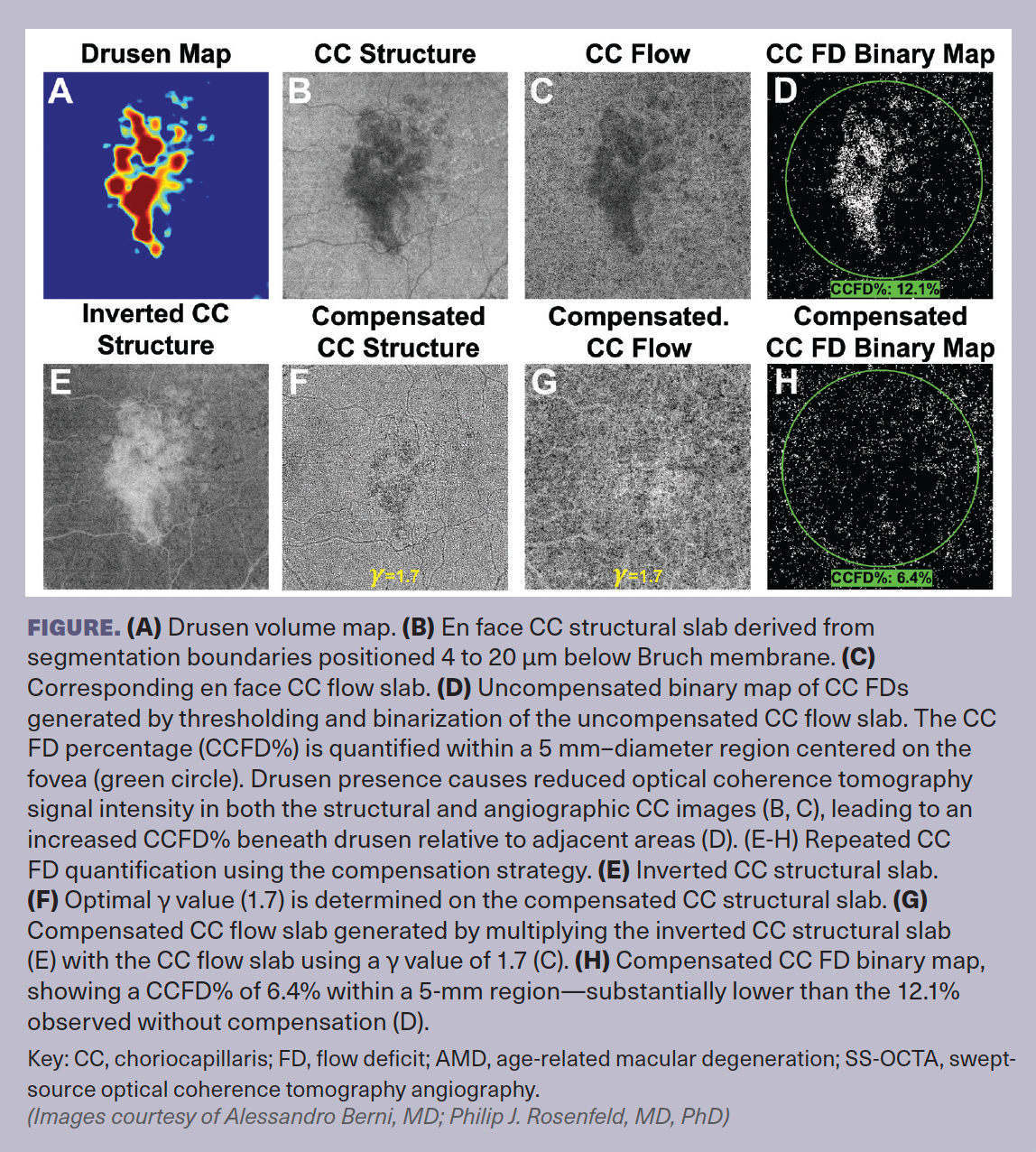
Recent advances in imaging techniques and compensation strategies have enhanced the accuracy of CC assessment in AMD.1,8-11 A key issue in CC imaging is the attenuation of OCT signal under drusen, which can create artificial increases in CC flow deficits (FDs). This artifact can lead to incorrect interpretations of CC perfusion status.

To address this, researchers have developed a compensation strategy that adjusts for signal loss, enabling more accurate quantification of CC FDs. By using en face structural CC images, this approach corrects attenuation artifacts and restores a more reliable flow signal. The method involves inverting the structural CC image and multiplying this latter by the CC flow image to enhance the OCTA signal in areas where it is decreased by drusen-induced attenuation.

Adjusting the inverted structural image

However, this simple multiplication is often insufficient to fully restore the signal under drusen, and subsequent observations highlighted the need for adapting the contrast of the inverted CC structural image by adjusting pixel intensity values using a power-law function. To address this limitation, we introduced a parameter, γ, which modifies the contrast by applying an exponential transformation to each pixel in the inverted structural image, raising its intensity to the power of γ. The γ parameter is greater than or equal to 1, and in most cases, a γ value between 1 and 2.5 provides an optimal balance between under- and overcompensation. This operation enhances signal compensation, allowing for greater correction in areas with severe attenuation while maintaining a balanced adjustment in regions with mild attenuation.

This compensation strategy is designed to account for the structural OCT signal loss that is apparent on CC structural slabs. Therefore, the appropriate level of compensation should be identified as the one that produces an even OCT signal across the CC structural slab. As such, the compensation strategy should first be applied to the CC structural image. However, the optimal level of compensation requires the use of objective approach to avoid over- or undercompensation.

We addressed this by analyzing the standard deviation (SD) of pixel intensities within the compensated structural image. The rationale behind this approach is that an evenly compensated CC structural image should exhibit a uniform signal distribution, minimizing variability across the slab. By testing different γ values and selecting the one that produces the lowest SD in pixel intensities, we can achieve an optimal correction tailored to each individual case. This approach ensures that OCT signal loss due to drusen-induced attenuation is adequately compensated, allowing for a more accurate assessment of CC FDs.

Consistent compensation across structural and flow images

Once the optimal γ value is determined based on the compensated structural image, this same γ value should be applied to compensate the CC flow image. This process ensures a consistent approach that provides an appropriate level of compensation for OCT signal loss, resulting in an optimally homogeneous level of OCT signal across the whole CC flow image. Consequently, any areas of decreased flow in the compensated CC flow image can be more reliably attributed to true flow deficits rather than artifacts from OCT signal loss.

This compensation strategy is shown in the Figure.

Challenges in imaging hypertransmission defects

Although compensation strategies have significantly improved CC imaging under drusen, other AMD-related lesions pose additional challenges. The retinal pigment epithelium (RPE) attenuates both incident and reflected light due to its scattering properties. In normal eyes and eyes with drusen, the compensation strategy described above is generally sufficient for CC imaging. However, when the RPE is thinned or absent, as in areas of geographic atrophy (GA), excessive light transmission into the choroid occurs, leading to what is termed choroidal hypertransmission defects (hyperTDs).12,13 These defects appear as regions of increased brightness on en face subRPE slabs (ranging from 64 to 400 µm beneath Bruch membrane) and are often associated with areas of focal hypopigmentation or GA as seen on color fundus imaging.

HyperTDs also affect en face CC slabs. The excessive light transmission results in bright regions on both CC structural and CC flow slabs, which, when inverted for compensation, appear darker. This inversion can lead to an artificial increase in CC FD measurements, making it appear as though perfusion is reduced in hyperTD regions when, in reality, the increased transmission simply alters the signal intensity.

To address this issue, we recommend either excluding hyperTDs from CC FD quantification or ensuring that they are not included in the compensation process. This can be achieved by identifying hyperTD areas on en face subRPE slabs, generating a mask, and applying this mask to the CC flow image before compensation. By doing so, the CC flow signal in these areas is preserved, preventing artificial CC FD elevations caused by excessive light transmission.

The impact of hypotransmission defects

Unlike hyperTDs, which result from increased light transmission, choroidal hypotransmission defects (hypoTDs) occur when OCT signal penetration is severely attenuated, rendering the CC undetectable. In AMD, these hypoTDs are primarily caused by HRF and CaD.7,8,14,15 HRF are well-defined lesions with a reflectivity equal to or higher than that of the RPE and are often linked to pigment migration into the retina. Their high scattering properties prevent OCT signal penetration wherever these lesions appear, leading to hypoTDs seen as intensely dark regions on the subRPE slabs. Similarly, CaD, which are drusen with a hyperreflective cap and heterogeneous internal reflectivity, scatter and absorb OCT light, preventing any meaningful imaging of the CC beneath them. Since hypoTDs represent areas where neither structural nor flow signals can be detected, they cannot be compensated.15 Attempting to apply compensation to these areas would yield unreliable CC FD measurements, as the missing signal does not represent true perfusion loss but rather an inability to image the CC.

To ensure accurate CC analysis, all hypoTDs should be identified and excluded from CC FD quantification. This can be done by generating masks based on subRPE slabs, similar to the approach used for hyperTDs, and applying these masks before binarization to prevent hypoTDs from influencing CC FD segmentation.

Longitudinal data further support the need for excluding hypoTDs. For example, in cases where HRF and CaD resolve over time, previously undetectable CC flow becomes visible again, and CC FD measurements return to levels similar to those in surrounding areas. This suggests that the high CC FD values previously observed in those regions were primarily due to the blockage of the OCT signal rather than true reductions in perfusion.

Accurate CC imaging in AMD requires a meticulous approach to ensure that observed flow deficits reflect true perfusion loss rather than imaging artifacts. Compensation strategies have proven effective in mitigating the effects of drusen-induced signal attenuation, but their application must be carefully implemented to prevent artefactual changes in CC FD measurements. Additionally, hyperTDs and hypoTDs pose significant challenges that require exclusion strategies to avoid miscalculation of the CC flow deficits.

Alessandro Berni, MD
E: Berni.Alessandro@hsr.it
Berni graduated from the University of Florence, Italy, in 2021 and is completing his ophthalmology residency at Vita-Salute San Raffaele University in Milan, Italy. From May 2023 to September 2024, he pursued a research fellowship in retinal diseases and advanced ophthalmic imaging at Bascom Palmer Eye Institute, University of Miami Miller School of Medicine in Florida, under the mentorship of Philip J. Rosenfeld, MD, PhD.
Philip J. Rosenfeld, MD, PhD
E: prosenfeld@med.miami.edu
Rosenfeld is a professor of ophthalmology at Bascom Palmer Eye Institute. He received research support from Carl Zeiss Meditec, Inc. Rosenfeld also received research funding from Novartis. He is also a consultant for AbbVie, Apellis, Bayer Pharmaceuticals, Boehringer-Ingelheim, Carl Zeiss Meditec, Character Biosciences, Genentech/Roche, InflammX Therapeutics, OcuDyne, Regeneron Pharmaceuticals, Sanofi, and Unity Biotechnology. He also has equity interest in Apellis, CharacterBio, InflammX, Ocudyne, and Valitor.
The remaining authors have no disclosures.
Research supported by grants from Carl Zeiss Meditec, Inc., an unrestricted grant from the Research to Prevent Blindness, Inc. (New York, New York), and the National Eye Institute (P30EY014801, R01 EY028753). The funding organizations had no role in the design or conduct of the present research.
References
1. Shi Y, Chu Z, Wang L, et al. Validation of a compensation strategy used to detect choriocapillaris flow deficits under drusen with swept source OCT angiography. Am J Ophthalmol. 2020;220:115-127. doi:10.1016/j.ajo.2020.06.033
2. Shi Y, Zhang Q, Zhou H, et al. Correlations between choriocapillaris and choroidal measurements and the growth of geographic atrophy using swept source OCT imaging. Am J Ophthalmol. 2021;224:321-331. doi:10.1016/j.ajo.2020.12.015
3. Iyer PG, Chu Z, Shen M, et al. Change in choriocapillaris flow deficits within tears of the retinal pigment epithelium imaged with swept-source optical coherence tomography angiography. Retina. 2022;42(11):2031-2038. doi:10.1097/IAE.0000000000003577
4. Li J, Liu Z, Lu J, et al. Decreased macular choriocapillaris perfusion in eyes with macular reticular pseudodrusen imaged with swept-source OCT angiography. Invest Ophthalmol Vis Sci. 2023;64(4):15. doi:10.1167/iovs.64.4.15
5. Zheng F, Zhang Q, Shi Y, et al. Age-dependent changes in the macular choriocapillaris of normal eyes imaged with swept-source optical coherence tomography angiography. Am J Ophthalmol. 2019;200:110-122. doi:10.1016/j.ajo.2018.12.025
6. Shi Y, Zhang Q, Zheng F, et al. Correlations between different choriocapillaris flow deficit parameters in normal eyes using swept source OCT angiography. Am J Ophthalmol. 2020;209:18-26. doi:10.1016/j.ajo.2019.09.017
7. Liu J, Laiginhas R, Shen M, et al. Multimodal imaging and en face OCT detection of calcified drusen in eyes with age-related macular degeneration. Ophthalmol Sci. 2022;2(2):100162. doi:10.1016/j.xops.2022.100162
8. Berni A, Shen M, Cheng Y, et al. The total macular burden of hyperreflective foci and the onset of persistent choroidal hypertransmission defects in intermediate AMD. Am J Ophthalmol. 2024;267:61-75. doi:10.1016/j.ajo.2024.06.023
9. Berni A, Kastner JD, Shen M, et al. Hyperreflective foci along the retinal pigment epithelium predict the onset of large choroidal hypertransmission defects in AMD. Am J Ophthalmol. Published online February 21, 2025. doi:10.1016/j.ajo.2025.02.021
10. Zhang Q, Zheng F, Motulsky EH, et al. A novel strategy for quantifying choriocapillaris flow voids using swept-source OCT angiography. Invest Ophthalmol Vis Sci. 2018;59(1):203-211. doi:10.1167/iovs.17-22953
11. Hiya FE, Cheng Y, Shen M, et al. A novel grid strategy for correlating focal macular anatomic changes with focal changes in choriocapillaris perfusion. Invest Ophthalmol Vis Sci. 2024;65(14):5. doi:10.1167/iovs.65.14.5
12. Shi Y, Yang J, Feuer W, Gregori G, Rosenfeld PJ. Persistent hypertransmission defects on en face OCT imaging as a stand-alone precursor for the future formation of geographic atrophy. Ophthalmol Retina. 2021;5(12):1214-1225. doi:10.1016/j.oret.2021.02.004
13. Laiginhas R, Shi Y, Shen M, et al. Persistent hypertransmission defects detected on en face swept source optical computed tomography images predict the formation of geographic atrophy in age-related macular degeneration. Am J Ophthalmol. 2022;237:58-70. doi:10.1016/j.ajo.2021.11.001
14. Laiginhas R, Liu J, Shen M, et al. Multimodal imaging, OCT B-scan localization, and en face OCT detection of macular hyperpigmentation in eyes with intermediate age-related macular degeneration. Ophthalmol Sci. 2022;2(2):100116. doi:10.1016/j.xops.2022.100116
15. Cheng Y, Hiya F, Li J, et al. Calcified drusen prevent the detection of underlying choriocapillaris using swept-source optical coherence tomography angiography. Invest Ophthalmol Vis Sci. 2024;65(6):26. doi:10.1167/iovs.65.6.26

Filed Under: Ophthalmology Times

Reader Interactions

Leave a Reply Cancel reply

Your email address will not be published. Required fields are marked *

Primary Sidebar

More to See

Week in Review: Patient Education Chatbot, Compensation Disparities Among Ophthalmology Faculty

April 3, 2026 By AAO News Feed

After a Failed Penetrating Keratoplasty, Either DSAEK or DMEK Could Be an Option

April 2, 2026 By AAO News Feed

Safety and Biodistribution of Topical Laquinimod, an Immunomodulator Targeting Aryl Hydrocarbon Receptor: The LION Study

April 2, 2026 By Ophthalmology Science

First-in-Human Clinical Evaluation of a Novel Non-Surgical Suprachoroidal Delivery Approach for Triamcinolone in Diabetic Macular Edema

April 2, 2026 By Ophthalmology Science

Who Gets Panretinal Photocoagulation? National Patterns in Proliferative Diabetic Retinopathy Treatment

April 2, 2026 By Ophthalmology Science

Weekly Journal Update — April 1, 2026

April 1, 2026 By AAO News Feed

A VR-Based Visual Field Test Has Utility in Children With Neuro-ophthalmic Disease

April 1, 2026 By AAO News Feed

Zero warm ischemia time in whole-eye transplantation through retrograde extracorporeal perfusion

April 1, 2026 By Ophthalmology Science

Some Patients With Punctate Inner Choroidopathy Can Experience Visual Impairment

March 31, 2026 By AAO News Feed

Patterns of X-linked Retinitis Pigmentosa Genetic Testing in England and Implications for Service Provision

March 31, 2026 By Ophthalmology Science

Footer

A RETINA-SPECIFIC ORGANIZATION

It’s clearly an advantage to work with a consulting group that’s focused on your industry. The team at Retina Consultant has over 50 years of retina-specific experience in healthcare business, practice management, marketing & operations.

We’ve got our eyes on working with the best retina groups and vendors in the industry.

See the best;  get in touch.

Recent News Posts

  • Tenecteplase Shows No Benefit Over Aspirin for Acute Central Retinal Artery Occlusion
  • Week in Review: Patient Education Chatbot, Compensation Disparities Among Ophthalmology Faculty
  • After a Failed Penetrating Keratoplasty, Either DSAEK or DMEK Could Be an Option
  • Safety and Biodistribution of Topical Laquinimod, an Immunomodulator Targeting Aryl Hydrocarbon Receptor: The LION Study
  • First-in-Human Clinical Evaluation of a Novel Non-Surgical Suprachoroidal Delivery Approach for Triamcinolone in Diabetic Macular Edema

Search

Retina Consultant
Copyright © 2026